How to Understand Your Mammogram Report

Lindsay Knake
| 3 min read
Lindsay Knake is a brand journalist for Blue Cross B...

Getting your mammogram is an important part of health for women age 40 and older.
Breast cancer, which affects about 13% of American women, is the second-leading cause of cancer-related deaths in U.S. women, according to BreastCancer.org.
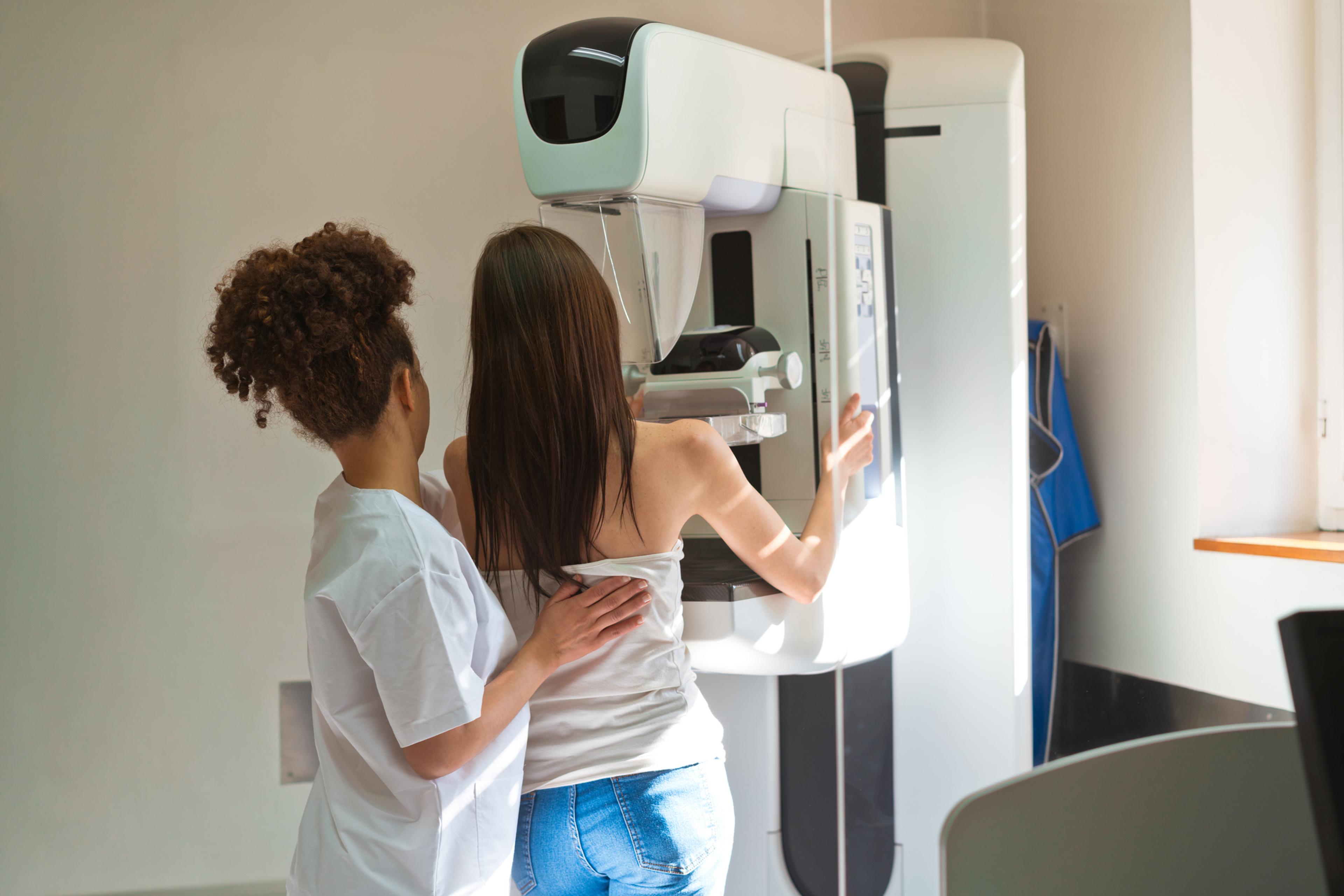
Mammograms are low-dose X-rays to check for changes in breast tissue that can indicate cancer. Women age 45-54 with an average risk for breast cancer should get annual screening mammograms and then screening mammograms every other year. Women who have higher risk of breast cancer should consult with their physician about when to start and how often to receive mammograms.
Your physician may order a diagnostic mammogram if you have symptoms of breast cancer.
What does a mammogram report include?
According to the American Cancer Society (ACS), radiologists use mammograms to check for:
- Calcification
- Masses
- Asymmetries
- Distortions
- Breast density
Calcifications
Calcifications are tiny calcium deposits in breast tissue that may or may not be caused by cancer. Macrocalcifications are larger and become more common with age. They are typically not cancerous. Microcalcifications are much smaller calcium deposits. A radiologist will determine if they look suspicious and need follow-up tests.
Masses
A mass is an area of abnormal breast tissue such as a cyst or tumor that may or may not be cancerous. Cysts are non-cancerous fluid-filled sacs. Solid masses are typically not cancerous, but your doctor may order follow-up scans to determine if the mass is cancer.
Asymmetries
Asymmetries are white areas in the mammogram that look different than typical tissue patterns that are typically not cancerous.
Distortions
Architectural distortions are an area of tissue that seems pulled toward a certain point, which could be a prior injury, positioning of the breast during the mammogram or a sign of cancer.
Breast density
Breast density is a measure of the amount of fibrous and glandular tissue compared to fatty tissue in your breast. About half of women have dense breasts, which has a higher risk of breast cancer. Dense tissue can make it more difficult to see abnormalities and cancer mammograms, but radiologists can still find cancer and abnormalities in mammograms.
As of September, the U.S. Food and Drug Administration (FDA) requires all mammogram reports include breast density, described in the report as “dense” or “not dense.”
What to look for in the mammogram results
After a mammogram, you will typically receive results in a few weeks. Your mammogram report will note if you have any abnormal findings and compare them to previous mammograms if you have one. Abnormal findings do not necessarily indicate you have breast cancer, but rather that your radiologist may seek follow up tests to determine whether the findings are cancerous or not.
The report gives results of mammograms with a score of 0 to 6 in the Breast Imaging Reporting and Data System (BI-RADS), according to the ACS.
- 0 indicates the mammogram is incomplete and you may an additional mammogram or further screenings.
- 1 indicates a normal test result with no abnormal findings.
- 2 indicates benign calcifications, masses, lymph nodes or changes in the breast.
- 3 indicates changes in the breast tissue are probably benign and a less than 2% chance of being cancer. Your physician may order a follow-up mammogram in 6 to 12 months to monitor these tissue changes until they are stable.
- 4 indicates a suspicious abnormality that may be cancer, and your physician will likely order a biopsy.
- 5 indicates the findings are highly suggestive of malignancy and have a 95% chance of being cancer. Your physician will likely order a biopsy.
- 6 indicates a known biopsy-proven malignancy. This category may be used in follow-up mammograms on breast cancer patients to see how treatment works.
Aside from annual physicals and mammograms, perform self-checks and report any changes to your doctor.
Image: Getty Images
Related:


